


A young woman feels a smooth, painless lump in her breast. Her doctor examines it, calls it a fibroadenoma, and tells her not to worry. Three months later, the lump is noticeably bigger. This is one of the most common reasons women in Punjab come in for a second opinion, and it is exactly where the question of phyllodes tumor vs fibroadenoma in Mohali becomes important. Both conditions can look almost identical at first glance, yet they behave very differently once growth speeds up.
Most rapidly enlarging breast lumps still turn out to be benign. But a growing lump deserves a closer look rather than quick reassurance, because separating a fibroadenoma from a phyllodes tumor in Mohali with certainty usually needs proper imaging and, often, a tissue sample.
Key Takeaways
- Fibroadenomas are the most common benign breast lumps in women under 35 and usually grow slowly.
- Phyllodes tumors are rare, making up roughly 0.3% to 1% of all breast tumors, but they tend to grow faster and reach a larger size than fibroadenomas.
- Rapid growth over weeks to a few months, not size alone, is the strongest clinical clue that pushes a doctor toward a phyllodes tumor rather than fibroadenoma.
- Ultrasound and mammography can suggest a diagnosis, but a core needle biopsy is usually needed to confirm it with confidence.
- Fibroadenomas rarely come back after removal, while phyllodes tumors need wider surgical margins because of a higher chance of local recurrence.
- Both conditions are managed successfully with timely diagnosis and the right surgical approach, so a growing lump is a reason to get checked, not a reason to panic.
Why Fibroadenoma and Phyllodes Tumor Look So Alike at First
Both lumps come from the same type of breast tissue. They belong to a group called fibroepithelial tumors, meaning they contain a mix of fibrous (connective) tissue and glandular tissue. That shared origin is exactly why a phyllodes tumor can feel and even scan like an ordinary fibroadenoma in its early stage.
On a first exam, both usually feel:
- Smooth or slightly lobulated
- Firm but not hard like a rock
- Painless
- Mobile under the fingers, not fixed to the skin or chest wall
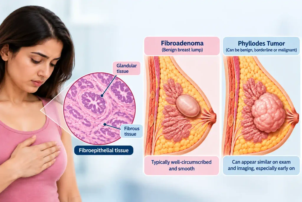
This overlap is not a diagnostic failure. It is simply biology. The real differences tend to show up over time, particularly in how fast the lump changes.
Fibroadenoma vs Phyllodes Tumor: The Differences That Matter
When comparing fibroadenoma vs phyllodes tumor, three factors carry the most clinical weight: growth rate, patient age, and what the tissue looks like under a microscope.
1. Growth Pattern and Speed
A typical fibroadenoma stays roughly the same size for months or years, or grows so slowly that the change is barely noticeable. A fast growing breast lump that visibly enlarges within a few weeks to a couple of months is far more suggestive of a phyllodes tumor, even if the earlier scan called it a fibroadenoma.
2. Age and Hormonal Pattern
Fibroadenomas are most common in the teens through the early thirties and are closely tied to estrogen activity. Phyllodes tumors typically appear later, most often between the ages of 40 and 50, although they can occur outside this range as well.
3. Feel and Texture on Examination
A fibroadenoma tends to feel uniformly smooth and rubbery. A phyllodes tumor, especially as it enlarges, can develop a firmer, multinodular, almost bulging quality, and the overlying skin may look stretched if the tumor is large.
| Feature | Fibroadenoma | Phyllodes Tumor |
| Growth speed | Slow, often stable for months or years | Often rapid, over weeks to a few months |
| Typical age group | Teens to early 30s | Usually 40 to 50 years |
| Texture on exam | Smooth, rubbery, freely mobile | Firmer, lobulated, sometimes multinodular |
| Average size noted at diagnosis | Around 2 to 3 cm | Often 4 cm or larger |
| Recurrence after removal | Uncommon | Higher, wider margins usually needed |
| Nature | Always benign | Benign, borderline, or malignant |
“A lump that keeps enlarging while everything else about it still looks fibroadenoma-like is exactly when I ask for tissue confirmation, not just another scan,” says Dr Balvin Kaur Ghai.
When a Fast-Growing Breast Lump Should Not Wait
Not every fast growing breast lump is a phyllodes tumor. Fibroadenomas can genuinely enlarge during pregnancy, before menstruation, or simply as part of normal hormonal fluctuation. So growth by itself is not proof of anything serious.
That said, certain patterns deserve a prompt visit rather than a wait-and-watch approach:
- The lump has visibly increased in size within the last one to three months
- A previous ultrasound called it a fibroadenoma, but it keeps growing anyway
- The lump feels different now than it did on first exam, firmer, lobulated, or less mobile
- There is skin stretching, visible bulging, or new discomfort over the area
- The lump is already larger than 3 to 4 cm at first detection
Any one of these on its own is not a red flag. Several together are a good reason to have the lump reassessed properly rather than assuming the earlier label still applies.
How Doctors Actually Tell Them Apart
1. What Ultrasound and Mammography Can (and Cannot) Show
Ultrasound is usually the first step for a breast lump in a younger woman, and it does a reasonable job of describing size, shape, and blood flow. Certain features on ultrasound, such as internal clefts or cystic spaces within a solid mass, can raise suspicion for a phyllodes tumor. Even so, imaging alone cannot reliably separate a phyllodes tumor from a fibroadenoma in every case, because the two can look nearly identical on a scan, particularly when the phyllodes tumor is still small.
2. Why a Tissue Biopsy Settles the Question
A core needle biopsy removes a small sample of tissue for examination under a microscope, and it remains the most dependable way to distinguish the two conditions. Pathology looks specifically at how the stromal (connective tissue) cells are arranged and how actively they are dividing, since this pattern is what separates a benign fibroadenoma from a fibroepithelial tumor with phyllodes features. For anyone weighing a breast biopsy Mohali option after a growing lump, this step is usually what finally brings clarity.
Getting Diagnosed in Mohali: What the Process Looks Like
A thorough workup for a growing breast lump generally follows three steps, and skipping any one of them can lead to a delayed diagnosis.
- Clinical examination, comparing growth rate, texture, and mobility against earlier notes
- Imaging, typically starting with ultrasound and adding mammography when needed
- Tissue sampling through a core needle biopsy, which confirms the diagnosis when imaging leaves any doubt
Access to a reliable ultrasound centre and a dependable pathology lab matters here, since biopsy results are only as trustworthy as the lab processing them. Our breast lump service covers this full pathway under one roof, from first exam through to confirmed diagnosis.
Treatment After Diagnosis
1. If It Turns Out to Be a Fibroadenoma
Small, stable fibroadenomas are often simply monitored. Larger or symptomatic ones, or those that keep growing, are usually removed through a straightforward excision, and recurrence at the same site is uncommon.
2. If It Turns Out to Be a Phyllodes Tumor
Surgical removal is standard, but the surgeon takes a wider margin of healthy tissue than a fibroadenoma excision would need. This matters because phyllodes tumors, even benign ones, carry a real chance of returning locally if margins are too narrow. Borderline or malignant cases may need further discussion around radiation, based on the pathology findings.
When to See a Breast Specialist in Mohali
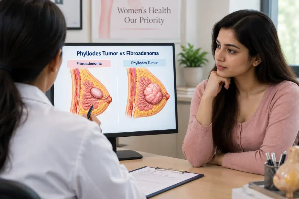
A growing breast lump does not need to become a source of ongoing worry, but it does deserve a proper look rather than repeated reassurance based on an old scan. Book an evaluation with a breast specialist Chandigarh or Mohali patients can reach easily if any of the following apply:
- A lump previously called a fibroadenoma is now clearly larger than before
- The lump has grown noticeably within the past few months
- You notice new firmness, skin changes, or discomfort over the area
- An earlier ultrasound left uncertainty about the diagnosis
- You simply want confirmation before deciding on monitoring versus removal
For a personal consultation on a growing lump, Dr Balvin Kaur Ghai’s clinical profile outlines her background in breast and gynaecological surgery for those who want to know more before booking.
Final Thought
Growth is information, not a verdict. A fibroadenoma that changes slowly is behaving exactly as expected, while a lump that enlarges quickly is simply asking for a second, closer look. Getting that look through proper imaging and, when needed, a biopsy is what turns uncertainty into a clear plan, whether that plan is watchful monitoring or a well-planned removal. Patients who address a growing lump early tend to have simpler surgery and far less anxiety along the way, and that outcome is achievable with the right diagnostic pathway in Mohali.
Frequently Asked Questions
1. What is the difference between a fibroadenoma and a phyllodes tumor?
Both are benign fibroepithelial breast lumps that arise from similar tissue, but a fibroadenoma is always non-cancerous and grows slowly, while a phyllodes tumor can grow quickly and, in a smaller proportion of cases, can be borderline or malignant. The main practical difference clinicians rely on is growth speed combined with tissue findings on biopsy.
2. Can a fibroadenoma grow quickly and still be benign?
Yes, fibroadenomas can enlarge noticeably during pregnancy, adolescence, or hormonal shifts and remain completely benign. Rapid growth raises the possibility of a phyllodes tumor, but it does not automatically rule out a fibroadenoma, which is exactly why reassessment rather than assumption matters.
3. How can doctors tell a fibroadenoma from a phyllodes tumor?
Doctors combine clinical examination, growth history, and imaging findings, then confirm the diagnosis with a core needle biopsy when there is any uncertainty. Tissue analysis under a microscope remains the most reliable method, since it directly examines how the stromal cells are behaving.
4. Does a fast-growing breast lump always mean it is a phyllodes tumor?
No, most fast growing breast lumps in younger women are still fibroadenomas responding to hormonal changes. Rapid growth simply lowers the threshold for further testing rather than confirming a phyllodes tumor on its own.
5. Can a phyllodes tumor look like a fibroadenoma on an ultrasound?
Yes, particularly when the phyllodes tumor is small, it can appear very similar to a fibroadenoma on ultrasound. Certain features, such as internal fluid-filled clefts, can raise suspicion, but ultrasound alone often cannot give a definitive answer.
6. What breast lump features make doctors consider a phyllodes tumor?
Rapid size increase, a firm or lobulated feel, size greater than 3 to 4 cm, and occurrence in women over 40 all raise the likelihood of a phyllodes tumor. None of these features alone is diagnostic, but several appearing together usually prompt a biopsy.
7. Can a fibroadenoma turn into a phyllodes tumor?
Most phyllodes tumors are thought to develop independently rather than transforming from an existing fibroadenoma, though a small number of case reports describe this kind of progression. Because this possibility exists, a fibroadenoma that changes character over time is worth reassessing rather than assuming it is still the same simple lump.
8. Is a breast biopsy needed to distinguish a fibroadenoma from a phyllodes tumor?
In most cases where growth or imaging findings raise any doubt, yes, a biopsy is the step that provides real confidence in the diagnosis. Without tissue confirmation, doctors are working from probability rather than certainty.
9. When should a growing fibroadenoma be checked again by a breast specialist?
Any noticeable increase in size over one to three months, or a change in how the lump feels, is a reasonable trigger to book a follow-up rather than waiting for the next routine check. Earlier reassessment generally leads to simpler management either way.
10. What type of biopsy is used when a phyllodes tumor is suspected?
A core needle biopsy is the standard first choice, since it removes enough tissue for the pathologist to assess the stromal pattern properly. In select cases, an excisional biopsy may be recommended if the core biopsy result remains inconclusive.
11. Where can I get a breast biopsy for a suspicious lump in Mohali?
Breast biopsy Mohali services are available through clinics that combine ultrasound-guided sampling with a dependable pathology partner. Our clinic coordinates this directly, from the initial scan through to result review, so patients are not left navigating multiple facilities alone.
12. When should I see a breast specialist in Chandigarh for a fast-growing lump?
If a lump has grown noticeably within a few months, feels different from before, or was previously diagnosed without a biopsy, that is a reasonable time to seek a specialist opinion rather than continuing to monitor informally. A short consultation is often enough to decide whether further testing is needed.
13. Does the size of a breast lump help distinguish a phyllodes tumor from a fibroadenoma?
Size is a supporting clue rather than a deciding one, since phyllodes tumors tend to be diagnosed at a larger average size than fibroadenomas. However, small phyllodes tumors and large fibroadenomas both exist, so size is always interpreted alongside growth rate and biopsy findings.
14. If an ultrasound says fibroadenoma but the lump keeps growing, what should I do?
Book a follow-up exam and consider a repeat ultrasound or biopsy rather than relying on the original report. An earlier scan reflects the lump as it was then, not necessarily how it behaves now.
15. How are phyllodes tumors and fibroadenomas treated after diagnosis in Mohali?
Fibroadenomas are either monitored or removed through a routine excision depending on size and symptoms, while phyllodes tumors are surgically removed with wider margins to reduce the chance of local recurrence. Both pathways are available locally, with the surgical approach tailored to the biopsy result and the individual patient.