

A breast lump that stays the same size rarely worries anyone for long. A breast lump that keeps growing is a different story, and it is the single most common reason women in Mohali book a consultation for what turns out to be a fibroadenoma. Growing fibroadenoma treatment in Mohali is not about rushing every lump into surgery. It is about knowing when watching and waiting stops being the safer choice.
Most fibroadenomas are harmless and never need anything more than periodic checking. But growth changes the conversation, because growth is one of the few features doctors cannot explain away with reassurance alone.
Key Takeaways
- Fibroadenomas are the most common benign breast lump, affecting up to 10% of women at some point in their lives.
- A typical fibroadenoma measures around 2 to 3 cm, while a “giant fibroadenoma” is defined as one larger than 5 cm.
- Growth, new pain, or a change in shape in a lump that was previously checked is a recognised reason to see a doctor again, not a reason to panic.
- Phyllodes tumors look and feel similar to fibroadenomas but behave differently, and only imaging or a biopsy can tell them apart.
- Removal is usually discussed case by case, based on size, growth rate, biopsy findings, and the woman’s own comfort with monitoring.
What Counts as a “Growing” Fibroadenoma?
Breast tissue changes throughout the month, and a little softness or tenderness before a period is normal. A growing fibroadenoma is different. It is a lump that gets measurably bigger over weeks or months, sometimes doubling in size, sometimes just creeping upward on repeat ultrasound scans.
Some fibroadenomas grow briefly during puberty or pregnancy and then settle. Others, called complex or juvenile fibroadenomas, tend to keep enlarging and are watched more closely. The pattern of growth, not the presence of a lump alone, is what shapes the next steps.
“I tell my patients the same thing every time: a lump that has not changed in a year behaves very differently from one that has doubled in three months. Both need attention, but only one usually needs a serious conversation about removal.” Dr Balvin Kaur Ghai
When Does a Growing Fibroadenoma Need Treatment?
Not every fibroadenoma that grows needs surgery, but certain patterns move the discussion from monitoring to active treatment. Doctors generally weigh a combination of factors rather than any single measurement.
- The lump has grown noticeably over two or more scans, not just felt slightly different on self-exam.
- It has crossed into the range where it starts distorting breast shape or pressing on surrounding tissue.
- Imaging or biopsy findings show features that are not entirely typical of a simple fibroadenoma.
- The lump is causing persistent discomfort, catching on clothing, or affecting confidence and daily life.
- The woman herself, after understanding her options, prefers removal over continued watching.
None of these on their own is an automatic trigger. A fibroadenoma close to 5 cm with stable, reassuring biopsy results may still be safely monitored in some cases, while a smaller one with unusual features on imaging might warrant earlier removal. This is exactly why growing fibroadenoma treatment in Mohali is planned around the individual case, not from a chart of measurements alone.
How Is a Growing Fibroadenoma Evaluated?
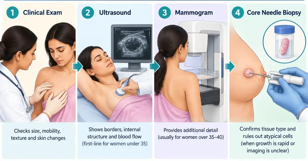
Evaluation usually starts with a clinical breast exam, where the doctor checks the lump’s size, mobility, texture, and any changes since the last visit. From there, imaging fills in details a physical exam cannot.
An ultrasound is typically the first step for women under 35, since breast tissue at this age is denser and ultrasound reads it more clearly than a mammogram. A good ultrasound centre will note the lump’s borders, internal pattern, and blood flow, all of which help separate a straightforward fibroadenoma from something needing closer inspection.
If anything on imaging looks atypical, or the lump has grown quickly, a core needle biopsy is usually recommended. This is a short outpatient procedure, and results from a reliable pathology lab generally confirm whether the tissue is a simple fibroadenoma, a complex variant, or something that needs a specialist’s further input.
| Evaluation Step | What It Checks | Typical Timing |
|---|---|---|
| Clinical exam | Size, mobility, tenderness, skin changes | At every visit |
| Ultrasound | Borders, internal structure, blood flow | First-line for women under 35 |
| Mammogram | Additional detail in denser or older breast tissue | Often added for women over 35–40 |
| Core needle biopsy | Confirms tissue type, rules out atypical cells | When growth is rapid or imaging is unclear |
Growing Fibroadenoma vs Phyllodes Tumor: What’s the Difference?
This is the question that brings the most anxious patients into the clinic, and it is a fair one. Fibroadenomas and phyllodes tumors form from similar breast tissue and can feel almost identical on examination. The difference lies in how they behave and how the cells look under a microscope.
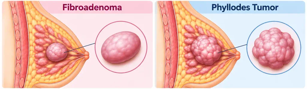
Phyllodes tumors tend to grow faster than typical fibroadenomas, and while most are benign, a small proportion can behave more aggressively. Research suggests phyllodes tumors account for less than 1% of all breast tumors, making them far rarer than fibroadenomas. Because the two conditions overlap so closely on a physical exam, imaging plus biopsy is the only reliable way to tell them apart.
| Feature | Fibroadenoma | Phyllodes Tumor |
|---|---|---|
| Growth speed | Usually slow, sometimes stable for years | Often faster, noticeable over weeks to months |
| Age group | Most common in the teens to mid-30s | Typically presents slightly later, often 30s to 50s |
| Feel on exam | Smooth, firm, rubbery, freely mobile | Can feel similar, sometimes firmer or lobulated |
| Behaviour | Almost always benign | Mostly benign, but a small subset can be malignant |
| Confirmed by | Ultrasound plus, if needed, biopsy | Core needle biopsy is essential, not optional |
| Typical management | Monitoring or removal based on growth and comfort | Surgical excision is usually recommended regardless of size |
The safest approach with any fast-growing lump is to treat the question seriously rather than guess from touch alone. A biopsy takes the uncertainty out of the equation.
When Fibroadenoma Removal May Be Discussed
Once evaluation is complete, removal becomes part of the conversation in a few recurring situations. A lump that has grown significantly since the last scan, one that is starting to affect the shape or comfort of the breast, or one where biopsy results raise any question mark are the usual candidates.
Removal is also often preferred by women who find repeated monitoring stressful, even when the lump itself is not medically urgent. That preference matters. Breast health decisions are not only clinical, they are personal, and a good breast lump surgery consultation should leave room for both the medical picture and how the patient feels about living with the lump.
Age and family history also shape the discussion. A growing lump in a woman with a strong family history of breast cancer is generally evaluated and, where appropriate, removed sooner than an identical lump in someone without that history, purely as a precaution.
What Fibroadenoma Removal Involves
Removal is typically done as a day-care procedure under local or short general anaesthesia, depending on the lump’s size and location. The surgeon makes a small incision, usually placed to minimise visible scarring, and removes the lump along with a thin margin of surrounding tissue.
Recovery for most women is quick. Mild soreness for a few days is normal, and most return to routine activities within a week, avoiding heavy lifting or strenuous exercise for a short period as the tissue heals. The removed tissue is always sent for pathology confirmation, even when imaging already looked reassuring, simply to close the loop with certainty.
Can a Fibroadenoma Grow Back After Treatment?
The lump that is removed does not grow back, since the tissue is physically gone. What can happen is that a new, separate fibroadenoma develops elsewhere in the same breast or in the other breast later on, because the underlying hormonal tendency that produced the first one has not changed.
This is why follow-up does not end at the stitches coming out. Periodic self-checks and, where recommended, follow-up imaging remain part of good breast health, regardless of whether a woman has had one fibroadenoma removed or several over the years.
Finding a Breast Lump Specialist in Mohali

Getting a growing breast lump assessed by the right clinician can make a real difference in deciding what happens next. Look for a specialist who examines the lump personally, reviews the actual ultrasound or biopsy images rather than relying only on the written report, and explains clearly why monitoring, further testing, or removal may be appropriate.
A trustworthy specialist will never push every patient toward surgery, and will equally never dismiss a genuinely growing lump with reassurance alone. You can read more about the background and approach of the team at CLAGS, or check general gynaecology surgery costs across Punjab before your visit, so the consultation focuses on your health rather than unfamiliar terminology.
Women considering a fibroadenoma removal, a fibroadenoma surgery in Chandigarh, or simply searching for a breast lump specialist in Mohali should expect the same starting point everywhere: a proper look at the lump before any recommendation is made.
Final Thought
A breast lump that grows deserves attention, not alarm. Most of the time, careful evaluation shows exactly what it is, a fibroadenoma behaving the way fibroadenomas sometimes do, and the path forward, whether that is watching or removing it, becomes clear once the facts are in hand. What matters most is not delaying that evaluation out of worry, and choosing a clinician who treats both the lump and the person carrying it with equal seriousness.
If you have noticed a breast lump that has changed or grown, book a consultation to get a clear, clinician-led answer rather than living with uncertainty.
Frequently Asked Questions
1. Does a growing fibroadenoma always need to be removed?
No, growth alone does not automatically mean surgery is required. Many fibroadenomas grow slowly and remain stable after evaluation, and monitoring with periodic ultrasound is often perfectly safe. Removal becomes more likely when growth is rapid, biopsy results raise questions, or the lump is affecting comfort or breast shape.
2. When should women in Mohali consider fibroadenoma removal?
Removal is usually considered when a lump has grown noticeably across two or more scans, when it is approaching a size that distorts the breast, or when a biopsy shows features that need closer management. It is also reasonable to consider removal simply because ongoing monitoring feels stressful, once the medical picture has been clearly explained.
3. Is a rapidly growing fibroadenoma a sign of cancer?
Rapid growth is more often linked to a complex fibroadenoma or a phyllodes tumor than to cancer, since typical fibroadenomas are almost always benign. That said, any lump that grows quickly should be evaluated with ultrasound and, if needed, a biopsy, because growth speed is one of the details doctors take seriously rather than ignore.
4. What causes a fibroadenoma to keep growing?
Fibroadenomas respond to hormonal changes, particularly oestrogen, which is why they can enlarge during puberty, pregnancy, or hormone therapy. Some fibroadenomas are simply biologically more active than others from the start, which is why growth patterns vary so much between women.
5. How is a growing fibroadenoma evaluated in Mohali?
Evaluation typically begins with a clinical breast exam followed by an ultrasound, which is the preferred first imaging test for younger women. If the lump looks atypical or has grown quickly, a core needle biopsy is added to confirm exactly what type of tissue is involved before any treatment decision is made.
6. Can ultrasound confirm whether a growing breast lump is a fibroadenoma?
Ultrasound gives strong clues, such as smooth borders and a typical internal pattern, that point toward a fibroadenoma rather than something more concerning. It cannot give a hundred percent tissue diagnosis on its own, which is why a biopsy is added whenever the picture is not completely clear-cut.
7. When is a biopsy recommended for a growing fibroadenoma?
A biopsy is generally recommended when the lump has grown quickly, when ultrasound features look even slightly atypical, or when the woman has risk factors such as a strong family history of breast cancer. It is a short outpatient procedure and remains the most reliable way to confirm tissue type.
8. How do doctors distinguish a growing fibroadenoma from a phyllodes tumor?
The two can feel almost identical during a physical exam, so doctors rely on ultrasound features and, most importantly, a core needle biopsy to tell them apart. Growth speed offers a clue, since phyllodes tumors often grow faster, but only tissue analysis gives a definite answer.
9. What size fibroadenoma usually requires removal?
There is no single size that automatically triggers surgery, though lumps approaching or exceeding 5 cm, sometimes called giant fibroadenomas, are removed more often simply because of the physical effect on breast shape. Growth pattern and biopsy findings usually matter more than the raw measurement alone.
10. What happens if a growing fibroadenoma is left untreated?
Many fibroadenomas can be left untreated safely with regular monitoring, especially when biopsy results are reassuring. If a genuinely growing or atypical lump is ignored entirely without follow-up, there is a risk of missing a change that needed earlier attention, which is why periodic review matters even during a watch-and-wait approach.
11. What does fibroadenoma removal involve?
Removal is usually a day-care procedure done under local or short general anaesthesia, where the surgeon takes out the lump through a small, carefully placed incision. Most women go home the same day and return to normal activities within about a week, with the tissue sent for pathology confirmation afterward.
12. How much does fibroadenoma surgery cost in Chandigarh or Mohali?
Cost depends on factors like the hospital, type of anaesthesia, and whether additional pathology testing is needed, so it genuinely varies between cases. A detailed, personalised estimate is usually given at consultation, and general regional pricing can also be reviewed on the gynaecology surgery cost page for context.
13. How can I find a breast lump specialist in Mohali for a growing fibroadenoma?
Look for a clinician who personally reviews your imaging, explains findings in plain language, and does not rush every lump toward surgery without cause. Checking a specialist’s background, patient reviews, and willingness to discuss both monitoring and removal honestly is a reasonable way to choose.
14. Can a fibroadenoma come back after surgical removal?
The exact lump that is removed cannot return, since the tissue itself is gone during surgery. A new, separate fibroadenoma can still develop later in either breast, because the hormonal tendency that caused the first one remains part of the body’s normal pattern.
15. Can a growing fibroadenoma be monitored instead of having surgery?
Yes, monitoring is a valid and common approach, particularly when growth is slow and biopsy results are reassuring. It usually involves repeat ultrasounds at set intervals, with surgery kept as an option if growth accelerates or the lump starts causing discomfort.